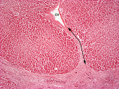
Cirrhosis (cirrhosis hepatis) 40x
The lobular structure of liver is totally disturbed. Regenerative nodi can be also seen in the sample. There are not any central veins in the middle of these lobules, it can be either asymmetrically located or totally missing. The amount of connective tissue has increased and it surrounds nodi. The connective tissue can be seen pink reaching from one portal area to another and also to the areas of the central vein (CV) (douple arrow). In cirrhotic liver there can be also gall stasis. Mild fatty degeneration is discovered. 40x magnification, HE-staining.


