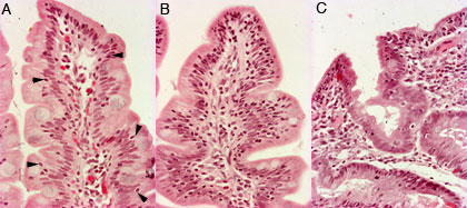
Atrophy of duodenal villi (atrophia villorum mucosae duodeni) 200x
There are three small intestine biopsies: normal mucosa (A), partial
villus atrophy (B) and subtotal villus atrophy (C). In the normal small
intestine the villi are thin and they form the major part, i.e. ¾ the
thickness of mucosa. The amount of lymphocytes and plasma cells in
lamina propria is normal. There are some lymphocytes in epithelium (arrowheds).
In partial villus atrophy villi are shorter and wider. There are plenty of lymphocytes in epithelium. THere are more cells in lamina propria, specially plasma cells.
In
subtoral villus atrophy changes are more dramatical than partial
atrophy. The mucosa seems to be flat. There is more hyperplasia in glands. The deformation of glands is obvious. 40x magnification, HE-staining.


 Systemic Pathology


