
level 1
Papillary Adenoma Sample |
|
Papillary Adenoma 40x A |
|
Papillary Adenoma 40x B |
|
Papillary Adenoma 200x A |
|
Papillary Adenoma 200x B |
|
Paksusuoli |
|
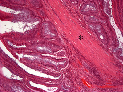
Papillary adenoma (adenoma villosum)
There can be seen a polypous tumor, which is expanding from the
intestinal mucosa. In the surface of tumor, villous structures are
seen. In the epithelium of villi nuclei are located in places in a
multilayer-way. There is mild dysplasia in this sample. Tumor is
restricted to the mucous membrane. The stem of tumor is made of
connective tissue (*). 40x magnification, HE-staining.